The DNase I amino acids fill the DNA minor groove, which becomes much wider than in canonical B-DNA. The relationship between the DNA backbone properties and protein binding via the minor groove was further demonstrated by probing the indirect readout mechanism underlying the DNA-DNase I interaction. The DNA of Nucleosome Core Particles alternates narrow and wide minor grooves associated with positive and negative rolls, in turn coupled with BI- and BII-rich regions, , respectively. Binding of the DNA minor groove by amino-acids is often accompanied by changes in ε and ζ. They are defined by the torsion angles ε and ζ, trans/g- in BI (ε-ζ∼−90°) and g-/trans in BII (ε-ζ∼+90°) ( Figure 1). These conformers were identified from crystallographic studies and by NMR. Also, the BI and BII conformational sub-states of phosphate groups appear associated with minor groove modulations in several DNA-protein complexes,. Hydrophobic contacts in the DNA minor groove could be aided by south to north sugar switches, north sugars increasing the accessibility of both sugars and bases. Thus, understanding the origin of minor groove widening remains a key question in structural biology, with far-reaching implications for DNA readout.Ĭrystallographic analyses of DNA-protein complexes suggested that conformational sub-states of the DNA phosphodiester backbone may be implicated in the minor groove binding mechanisms. This interesting finding helps to understand how proteins can penetrate into a narrow DNA minor groove but does not explain the mechanisms underpinning the enlargement of the minor groove in many bound DNAs, which can be considerable.
DNA BACKBONE FREE
T rich segments that tend to adopt very narrow minor grooves in both free and bound DNA crystal structures.This electrostatic effect is particularly marked in A Poisson-Boltzmann calculations revealed that narrow minor grooves exhibit an enhanced negative electrostatic potential, favoring their interaction with arginine residues. This view has been recently revisited in a study addressing very large datasets of free and bound X-ray DNA structures, showing that the electrostatic potential in the minor groove is influenced by its geometry, and can be recognized by proteins. Nevertheless, this type of interaction is still intriguing since the DNA minor groove is often presumed too narrow to accommodate protein structural elements without energetically costly distortions. Architectural proteins and proteins binding DNA sequences non-specifically mainly interact with the DNA minor groove, where there is little discrimination between base types. More contacts than expected are observed in the DNA minor groove. Indeed, the increasing number of X-ray structures highlighted the importance of DNA minor groove in DNA-protein complexes. The major groove dimensions remain quite similar in free and bound DNA in contrast, the minor groove is especially variable in complexes. Thus, sequence specific variations of the DNA grooves play a central role in DNA-protein readout processes –. In DNA-protein complexes, proteins fit snugly in the DNA major and minor grooves. Therefore, understanding the origins of the conformational preferences of the free nucleotidic DNA sequences remains an important goal in structural biology. The DNA binding process depends on the intrinsic ability of free DNA to adopt its structure when bound to a protein. DNA-protein interactions are informed by the intrinsic mechanical properties of DNA, which facilitate its deformation in the complex.

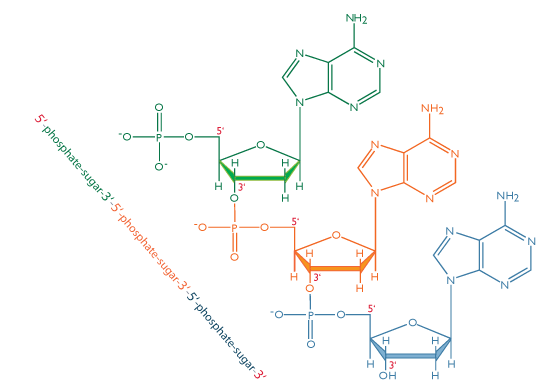
Stryer L.The cellular DNA is continuously “read” by proteins. Kilpatrick S.T 2011 Lewin's Genes X, 10th Edition, Jones and Bartlett Publishers: London These two sugars only differ by one -OH group being changed to an -H, but provides different capabilities for each molecule. On on the other hand, the sugar in the backbone of RNA is called ribose. In DNA, the sugar involved is deoxyribose. However, their sugar phosphate backbone differs slightly. RNA and DNA are both examples of phosphodiesters and have a very similar structure. One turn of this helix is 34nm long, the diameter of it is 2nm, and there are ten bases attached per turn at 0.34nm. These features make DNA can repel water and would not hydrolysed and breakdown by the aqueous environment.

DNA is very stable due to rungs of “ladder” is hydrophobic and phosphate sugar backbone of DNA is negatively charged. The purpose of this twisting is to protect the bases inside it, and prevent them from being damaged by the environment. one runs 3' to 5', the other run 5' to 3'. This is done by the sugar phosphate backbone twisting around itself in a coil. Figure 1 Diagram showing the sugar phosphate backbone of DNA, and the nitrogenous bases attached to it, forming a nucleotide Structure of DNAĭNA is wound into an right-handed double helix.


 0 kommentar(er)
